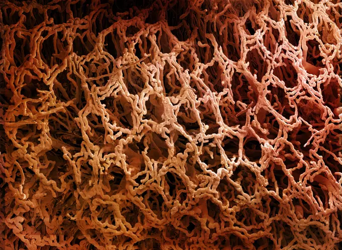
Colorized scanning electron micrograph of Marburg virus particles both budding and attached to the surface of an infected VERO E6 cell (gold).
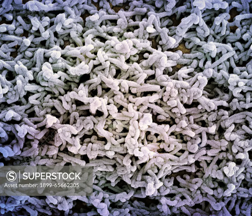
SuperStock offers millions of photos, videos, and stock assets to creatives around the world. This image of Colorized scanning electron micrograph of Marburg virus particles both budding and attached to the surface of an infected VERO E6 cell (gold). by NIH-NIAID/IMAGE POINT FR/BSIP/Universal Images is available for licensing today.
Visually Similar More from Martian Terrain and Fossils story


Looking for a license?
Click here, and we'll help you find it! Questions? Just ask!
Click here, and we'll help you find it! Questions? Just ask!
DETAILS
Image Number: 1899-65662305Rights ManagedCredit Line:NIH-NIAID/IMAGE POINT FR/BSIP/Universal Images/SuperStockCollection:Universal Images Story:Martian Terrain and FossilsContributor:NIH-NIAID / IMAGE POINT FR / BSIP Model Release:NoProperty Release:NoResolution:4096×3509