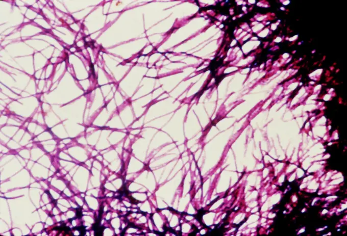
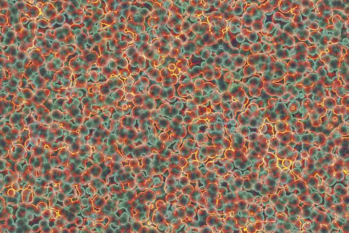
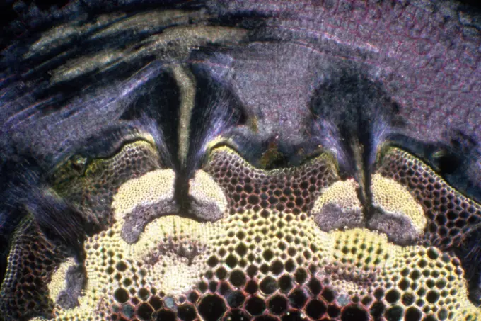
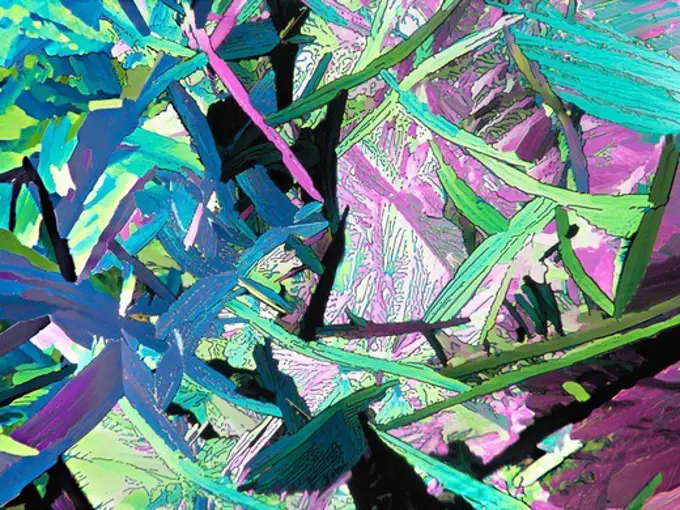
This award-winning image of the hippocampus with an ultra-wide, high-speed multi-photon laser microscope. The tissue was stained to reveal the organization of glial cells (cyan), neurofilaments (green) and DNA (yellow). The microscope used by Deerinck was developed in collaboration with Roger Tsien (Nobel Prize in Chemistry 2008) and remains a powerful and unique tool today (2012, almost 20 years after its development).
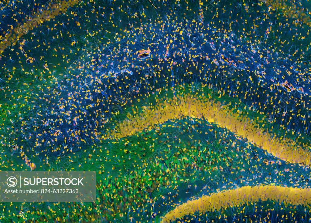
SuperStock offers millions of photos, videos, and stock assets to creatives around the world. This image of This award-winning image of the hippocampus with an ultra-wide, high-speed multi-photon laser microscope. The tissue was stained to reveal the organization of glial cells (cyan), neurofilaments (green) and DNA (yellow). The microscope used by Deerinck was developed in collaboration with Roger Tsien (Nobel Prize in Chemistry 2008) and remains a powerful and unique tool today (2012, almost 20 years after its development). by NIH/IMAGE POINT FR/BSIP is available for licensing today.
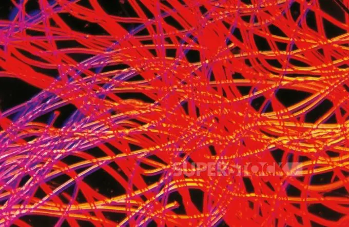
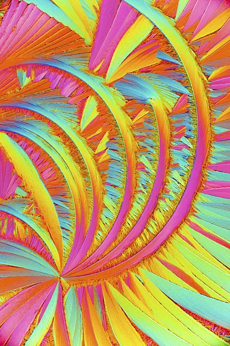
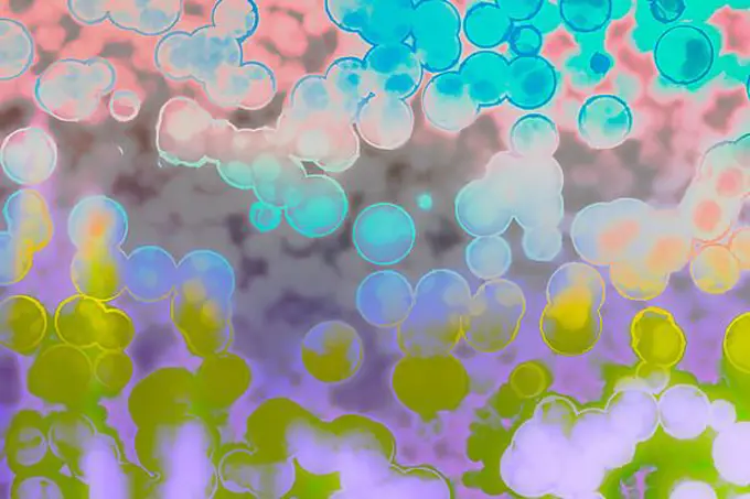
Visually Similar More from Microscopic Wonders story
Looking for a license?
Click here, and we'll help you find it! Questions? Just ask!
Click here, and we'll help you find it! Questions? Just ask!
DETAILS
Image Number: 824-63227363Rights ManagedCredit Line:NIH/IMAGE POINT FR/BSIP/SuperStockCollection:BSIP Story:Microscopic WondersContributor:NIH / IMAGE POINT FR / BSIP Model Release:NoProperty Release:NoResolution:4204×3020