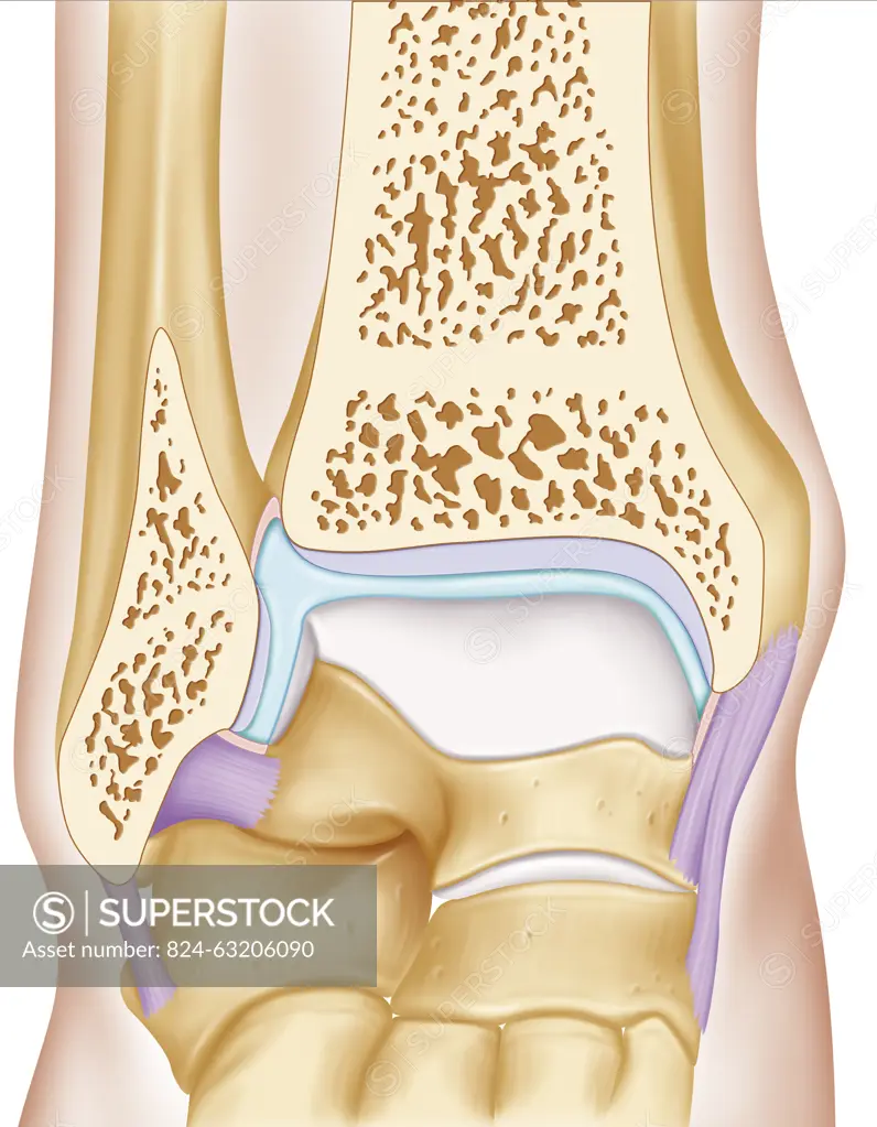
Anterior view illustration of the ankle joint. The two joint surfaces (grey) are visible, the joint surface of the fibula and tibia distal epiphysis and that of the talus. The articular capsule is filled with synovial fluid (blue) surrounded by its membrane (pink), it is attached to the lateral ligaments.
SuperStock offers millions of photos, videos, and stock assets to creatives around the world. This image of Anterior view illustration of the ankle joint. The two joint surfaces (grey) are visible, the joint surface of the fibula and tibia distal epiphysis and that of the talus. The articular capsule is filled with synovial fluid (blue) surrounded by its membrane (pink), it is attached to the lateral ligaments. by JACOPIN/BSIP is available for licensing today.
Looking for a license?
Click here, and we'll help you find it! Questions? Just ask!
Click here, and we'll help you find it! Questions? Just ask!
DETAILS
Image Number: 824-63206090Rights ManagedCredit Line:JACOPIN/BSIP/SuperStockCollection:BSIP Contributor:JACOPIN / BSIP Model Release:NoProperty Release:NoResolution:3950×5079