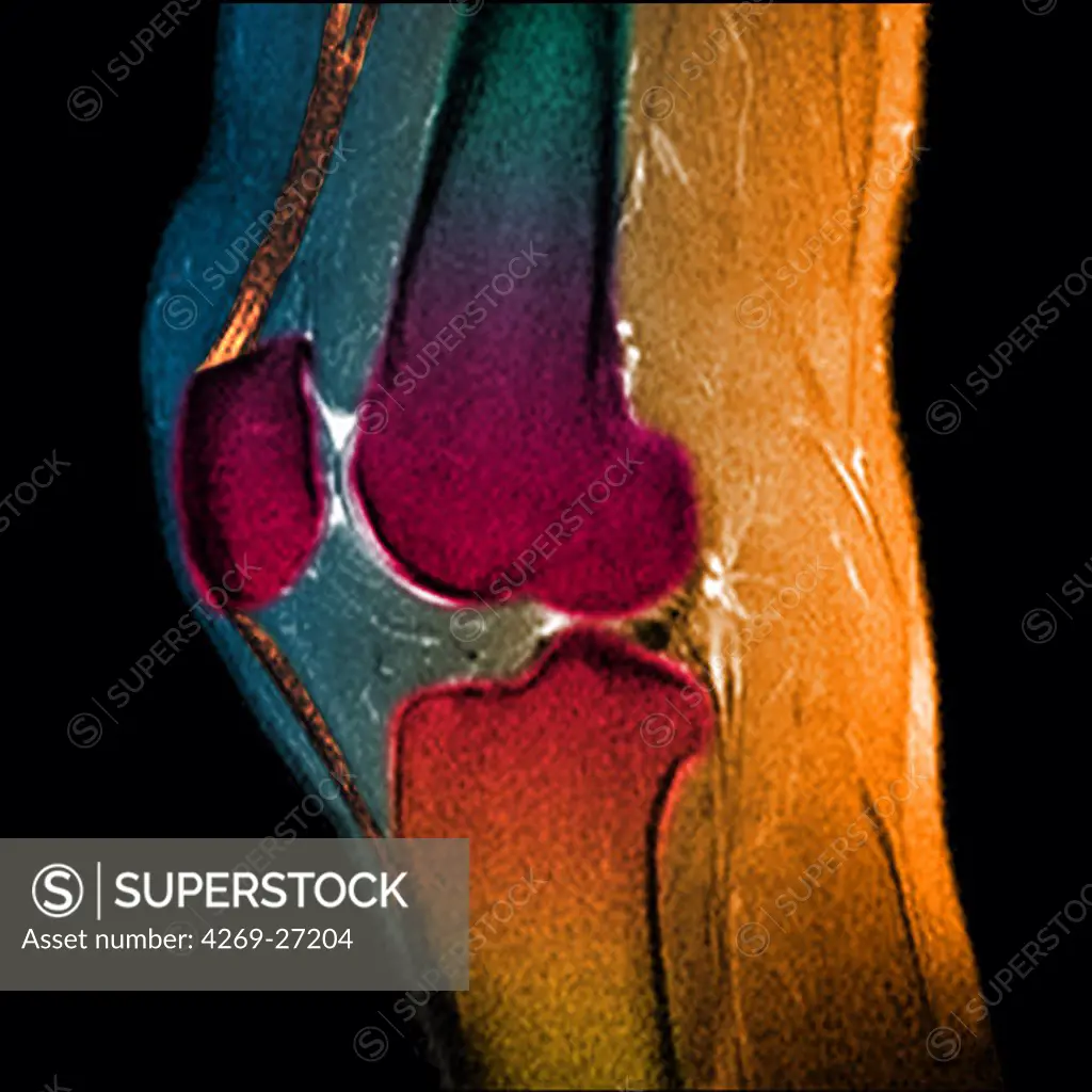
Knee. Sagittal (side view) Magnetic Resonance Image (MRI) of the knee showing in orange the patellar tendon (between the patella and the tibia above), and the flexing quadriceps tendon (between the patella and the femur below). The distal femur (above), the proximal tibia (below), and the patella or knee cap (left) are in red.
SuperStock offers millions of photos, videos, and stock assets to creatives around the world. This image of Knee. Sagittal (side view) Magnetic Resonance Image (MRI) of the knee showing in orange the patellar tendon (between the patella and the tibia above), and the flexing quadriceps tendon (between the patella and the femur below). The distal femur (above), the proximal tibia (below), and the patella or knee cap (left) are in red. by Guilloz-Chu Nancy/Phanie is available for licensing today.
DETAILS
Image Number: 4269-27204Rights ManagedCredit Line:SuperStock / Guilloz-Chu Nancy/PhanieCollection: Phanie Contributor: Guilloz-Chu Nancy Model Release:NoProperty Release:NoResolution:4500×4500
Free Research
Can't find the usage you need?
We're here to help!
Phone: +1 866 236 0087
Email: help@superstock.com
Research / License Request Form
Can't find the usage you need?
We're here to help!
Phone: +1 866 236 0087
Email: help@superstock.com
Research / License Request Form
Retouching Services
Our MediaMagnet division offers comprehensive retouching services at great rates. For a free quote, please send us an e-mail and we'll get back to you promptly.
Email: Retouching Service
Our MediaMagnet division offers comprehensive retouching services at great rates. For a free quote, please send us an e-mail and we'll get back to you promptly.
Email: Retouching Service